


North Texas GME Research Forum 2024
Files
Download Poster or Presentation (5.2 MB)
Division
North Texas
Hospital
Medical City Fort Worth
Specialty
Internal Medicine
Document Type
Poster
Publication Date
2024
Keywords
Leukocytoclastic Vasculitis, purpura
Disciplines
Internal Medicine | Medicine and Health Sciences | Skin and Connective Tissue Diseases
Abstract
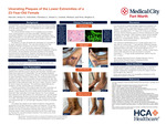
INTRODUCTION: What should come to mind when any physician sees dark red spots on the legs? If you answered vasculitis, then you are on the right track. Leukocytoclastic vasculitis (LCV) is defined as small vessel inflammation of the dermal capillaries and venules. The clinical hallmark finding of LCV is palpable purpura (raised, round, purple plaques) on the lower extremities. LCV is confirmed by histopathology showing neutrophilic infiltration in the walls of dermal vasculature. Though LCV is the most common cause of clinical vasculitis, the annual incidence of biopsy-proven LCV is approximately 30-45 per million individuals. LCV typically occurs in adults and has no gender predilection. Though idiopathic 60% of the time, LCV may be secondary to other causes, including infection, drugs, systemic disease, or malignancy. CASE PRESENTATION: We present a 23-year-old African-American female who developed bilateral lower extremity purpuric plaques with secondary ulceration and bullae three weeks prior to presentation. Her skin findings manifested several days following a camping trip to Mexico, though she also endorsed an upper respiratory infection 6 weeks prior. She previously sought medical treatment on two other occasions and had been prescribed a 9 day prednisone taper and naproxen. The prednisone helped alleviate her symptoms, but they returned after she completed the taper. Biopsies were obtained with histopathology showing a perivascular neutrophilic infiltrate consistent with LCV. The patient was started on four weeks of systemic corticosteroids (prednisone 20 mg daily followed by a taper) and dapsone 100 mg daily. NSAIDs and hydrocodone were initiated for pain control as needed. Leg elevation and use of compression stockings were utilized for lower extremity edema. Patient showed improvement at the three week follow up, and demonstrated resolution of her ulcers at six-month follow-up. LEARNING POINTS: Many cases of LCV are self-limited and resolve after weeks to months. Treatment depends on etiology and disease severity. This case demonstrates how initial suboptimal treatments can result in progression to severe/chronic LCV. In patients with chronic disease duration and/or complicated physical exam findings (eg, ulceration/bullae), it is important to initiate at least 4 weeks of systemic steroid therapy as soon as possible. Subsequent management involves the usage of steroid-sparing agents (eg, dapsone). It is valuable that the primary care physician and general hospitalist recognize this condition, as early detection and timely treatment prevents serious complications.
Original Publisher
HCA Healthcare Graduate Medical Education
Recommended Citation
Okereke, Robyn O.; Scheufele, Christian J.; Carletti, Michael; Weis, Stephen E.; and Bahrami, Carlos, "Ulcerating Plaques of the Lower Extremities of a 23-Year-Old Female" (2024). North Texas GME Research Forum 2024. 38.
https://scholarlycommons.hcahealthcare.com/northtexas2024/38