


North Texas GME Research Forum 2024
Files
Download Poster or Presentation (749 KB)
Division
North Texas
Hospital
Medical City Fort Worth
Specialty
Dermatology
Document Type
Poster
Publication Date
2024
Keywords
skin pigmentation, tinea corporis, ringworm
Disciplines
Bacterial Infections and Mycoses | Dermatology | Medicine and Health Sciences | Skin and Connective Tissue Diseases
Abstract
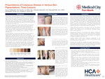
Introduction: Tinea corporis, a superficial fungal skin infection, typically manifests as pruritic annular erythematous scaly plaques with central clearing. This condition can involve every body region. Here we present tinea corporis across various skin pigmentations. The goal of this presentation is to highlight distinctions and similarities in light, medium, and dark skin tones, using the Fitzpatrick scale for stratification of skin types. We hope this can aid primary care clinicians in early recognition of this common condition. Case Presentation: We present a series of cases illustrating the diverse manifestations of tinea corporis across different Fitzpatrick skin types. In Figure 2, a 28-year-old with Fitzpatrick I displays well-defined, scaly plaques on the inner thigh. Figure 3 features a 3-year-old of Fitzpatrick II with a large, poorly defined plaque on the right posterior thigh. A 33-year-old Fitzpatrick III male of showcases an erythematous, scaly plaque extending from the groin (Figure 4). Figure 5 presents a 15-year-old with Fitzpatrick IV, exhibiting well-demarcated erythematous plaques on the left arm with inflamed papules on the neck and arm. A 69-year-old female of Fitzpatrick V displays pink to violaceous plaques with hyperpigmentation and white/gray scales (Figure 6). These cases highlight the diverse clinical presentations that can be seen in different Fitzpatrick skin types. Learning Points: Tinea corporis is a common superficial fungal infection of the skin that tends to be more prevalent in younger demographics and thrives in humid environments. Trichophyton rubrum is the most common dermatophyte culprit for this fungal infection. It can spread easily between different parts of the body, accentuating the need for comprehensive skin examination encompassing all anatomical areas. Arriving at the correct diagnosis of tinea corporis across various skin tones involves recognizing the influence of age, prior and concomitant treatments, environmental factors, and the Fitzpatrick scale on its clinical presentation. Variations can be observed in the degree of scaling and the amount of erythema. Erythema is difficult to identify in darker Fitzpatrick skin types (IV-VI). Instead, erythema may appear hyperpigmentation. Likewise, scale may be more easily seen in these skin types. Identifying tinea corporis in light, medium, and dark skin tones is crucial for early intervention. Confirmatory diagnostic procedures, such as KOH scrapings and fungal cultures from lesion swabs are valuable in confirming the presence of dermatophyte involvement. If clinical suspicion remains high, a skin biopsy can be obtained for histologic confirmation.
Original Publisher
HCA Healthcare Graduate Medical Education
Recommended Citation
Peddireddy, Navya; van Alfen, Braden; Scheufele, Christian; Hall, Marshall; Wong, Christopher M.; and Weis, Stephen, "Presentations of Cutaneous Disease in Various Skin Pigmentations: Tinea Corporis" (2024). North Texas GME Research Forum 2024. 47.
https://scholarlycommons.hcahealthcare.com/northtexas2024/47
Included in
Bacterial Infections and Mycoses Commons, Dermatology Commons, Skin and Connective Tissue Diseases Commons