

Division
East Florida
Hospital
HCA Florida Westside Hospital
Specialty
Pathology
Document Type
Poster
Publication Date
2023
Keywords
medulloblastoma, tumor, brain tumor
Disciplines
Medicine and Health Sciences | Neoplasms | Nervous System Diseases | Pathology
Abstract
Introduction: Medulloblastoma (MB) is an embryonal central nervous system (CNS) tumor located in the posterior cranial fossa. Medulloblastoma is one of the most common malignant pediatric brain tumors, accounts for 25% of all pediatric intracranial tumors, but rare in adults; and constitute 0.4%-1% of adult primary brain tumors. The incidence of medulloblastomas in adults is approximately 0.5 per million per year and decreases with age with male predominance. Medulloblastomas classically appear as well-defined masses in the cerebellum that enhance both computed tomography (CT) of the head and brain magnetic resonance imaging (MRI) with gadolinium contrast. The clinical symptoms and signs of MB in adults are associated with the location of the tumor, increased intracranial pressure, and/or obstruction of the cerebrospinal fluid pathway leading to headache, dizziness, nausea, ipsilateral cerebellar signs, and ataxia.
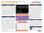
Case Presentation: A 31-year-old female presented with 3 weeks of intermittent headaches with dizziness, followed by vomiting. The neurological exam was unremarkable. The patient was started on dexamethasone after a non-contrast brain CT revealed a 5 cm ill-defined cerebellar mass, and was suggestive of early herniation. Follow-up magnetic resonance imaging with contrast showed a 4.7x4.1x4.3 cm enhancing extra-axial mass. Histopathologic evaluation of the tumor biopsy showed a highly cellular embryonal neoplasm with prominent nuclear molding and brisk mitotic activity, and also scattered karyorrhexis; and occasional areas of the tumor show apparent nodular growth. Some tumor cells were immunoreactive for GFAP while synaptophysin shows weak reactivity in much of the tumor with slightly stronger staining of tumor nodules. A reticulin-rich stroma is apparent in many areas. Molecular studies showed p53 wild-type pattern with IHC, no amplification of MYC by FISH, no amplification of MYCN by FISH, SHH molecular group. This case is diagnosed as Medulloblastoma, desmoplastic/nodular, SHHactivated, CNS WHO grade 4.
Discussion: In a study done by Zhao et al., it was shown that there is a preponderance of SHH-type tumors in adult MB (62%), followed by group 4 tumors (28%) and WNT-activated tumors (10%), with an absence of group 3 cases, suggesting that this subgroup may be restricted to pediatric MB. There have been only 12 reported cases of extra‑axial MB in the adult literature. There have been only 12 reported cases of extra‑axial MB in the adult literature.
Recommended Citation
Wymer, Gul; Mousavi, Fatemeh; and Barker, Jennifer, "Extra-axial Desmoplastic/Nodular Medulloblastoma, SHH-activated in adult: a case report" (2023). East Florida Division GME Research Day 2023. 40.
https://scholarlycommons.hcahealthcare.com/eastflorida2023/40