


North Texas GME Research Forum 2026
Files
Download Full Text (588 KB)
Division
North Texas
Hospital
Medical City Fort Worth
Specialty
Internal Medicine
Document Type
Poster
Publication Date
2026
Keywords
jejunal arteriovenous malformation, AVM
Disciplines
Congenital, Hereditary, and Neonatal Diseases and Abnormalities | Gastroenterology | Internal Medicine | Medicine and Health Sciences
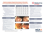
Abstract
Small bowel bleeding is relatively uncommon, accounting for less than 10% of all gastrointestinal (GI) bleeds. Small bowel angiodysplasias are the most common cause of small bowel bleeding. These lesions can often be difficult to identify on radiographic imaging or endoscopy and tend to rebleed after intervention. Our case centers on an 86-year-old patient who presented with an acute GI bleed from an obscure ulcerated jejunal arteriovenous malformation (AVM); his past medical history includes an undisclosed prior upper GI bleed treated with endoscopic clipping, paroxysmal atrial fibrillation, type 2 diabetes, prostate cancer with prostatectomy, and other chronic conditions. He presented for evaluation with symptomatic anemia from melenic stools. On admission, hemoglobin and hematocrit were 6.7g/dL and 22.9%, requiring 2 units of packed red blood cells to be transfused. Patient continued to have melena with persistent drops in hemoglobin throughout this admission requiring a total of 25 units of packed red blood cells along with IV iron infusions. The patient underwent small bowel enteroscopy (SBE) and Colonoscopy. SBE was grossly unremarkable. Colonoscopy revealed old blood but no active source of bleeding. Video capsule endoscopy (VCE) was performed which revealed trace amounts of old blood in the proximal small bowel with no clear etiology. A Tagged red blood cell scintigraphy was performed and determined that the source of bleeding was in the jejunum. A provocative CT angiogram was performed to localize and embolize the source however it was unsuccessful as there was no active extravasation noted during the study. Due to failed radiographic approaches to manage the patient’s ongoing bleed a repeat SBE beyond the extent of previous endoscopic reach was performed. The repeat SBE was notable for multiple jejunal arteriovenous malformations (AVMs), one of which was large and ulcerated and likely the etiology of notable blood loss requiring multiple blood transfusions. Argon plasma coagulation (APC) was performed on the smaller AVMs with successful ablation. APC along with endoclipping was performed on the ulcerated AVM resulting in successful ablation. The patient demonstrated rapid clinical improvement with resolution of melena, confirmed with a normalized hemoglobin, and he was eventually discharged. This case highlights the unique difficulty in diagnosing and managing an obscure type of small bowel bleed from ulcerated angiodysplastic lesions, as these produce greater blood loss but remain difficult to identify on mesenteric imaging and different types of endoscopy.
Original Publisher
HCA Healthcare Graduate Medical Education
Recommended Citation
Mathews, Joel; Lewis, Trina; Mathew, John; and Hoang, Long, "Negative Imaging, Ongoing Hemorrhage: An Ulcerated Jejunal AVM Requiring Advanced Endoscopic Intervention" (2026). North Texas GME Research Forum 2026. 12.
https://scholarlycommons.hcahealthcare.com/northtexas2026/12
Included in
Congenital, Hereditary, and Neonatal Diseases and Abnormalities Commons, Gastroenterology Commons, Internal Medicine Commons