

Division
East Florida
Hospital
HCA Florida Aventura Hospital
Specialty
Pulmonary Critical Care
Document Type
Poster
Publication Date
2023
Keywords
marijuana use, vanishing lung syndrome, VLS, idiopathic giant bullous emphysema
Disciplines
Critical Care | Internal Medicine | Medicine and Health Sciences | Respiratory Tract Diseases
Abstract
Please see supplemental content for full abstract with figures and references.
Introduction: Vanishing Lung Syndrome (VLS), also known as idiopathic giant bullous emphysema, is a rare pulmonary condition characterized by the development of large, thin-walled bullae within the lung parenchyma. While the exact etiology of VLS remains unclear, there is growing interest in exploring a potential association between VLS and cannabis use. This case report aims to contribute to the existing body of knowledge by presenting a case of VLS in a patient with a history of chronic cannabis consumption in prior years.
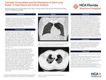
Clinical Presentation: A 44-year-old former smoker male with a history of mild intermittent asthma and marijuana use presented to the hospital with concerns of shortness of breath and a non productive cough. Patient reports symptoms started one day prior to presentation and were associated with subjective fevers, chills, wheezing, and pleuritic chest pain. The dyspnea was described as constant, on rest, exacerbated by exertion, and partially relieved with albuterol inhaler. Physical exam was significant for wheezing in bilateral lung fields and decreased breathing sounds in upper lobes. A computed tomography of the chest with contrast showed bilateral massive bullae in the upper 40% of the right lung and slightly larger size in the left lung (Figure 1,2).
Pulmonary function tests at rest, performed after hospital discharge, were significant for a decreased FEV1/FVC ratio and normal DLCO consistent with asthma disorder. Alpha antitrypsin levels were within normal limits as well as autoimmune workup. Patient was referred to the cardiothoracic surgeon who intraoperatively found a giant bulla approximately 15 cm involving the anterior segment and compressing the remaining of the left lung, with significant adhesions to the pericardium, and a 10 cm bulla involving the apical portion of the posterior apical segment. As a result, it was performed a segmentectomy of a portion of the apical posterior lobe, an anterior lobe segmentectomy, lysis of the adhesions, and excision of the bulla from the superior segment. Bullous emphysema was confirmed histologically, in addition to mild chronic inflammation, vascular congestion, and focal organizing pneumonia-like features. Patient was discharged home without need for home oxygen and symptoms improvement.
Discussion: VLS is a rare lung condition which typically affects young male smokers. Bullous emphysema refers to emphysematous lung with bullae, which are air-filled spaces within the parenchyma that are 1 cm or larger in diameter and consist of a thin wall of visceral pleura with remnants of alveolar and interlobular septa inside.(1) The most common associated risk factor to developing this condition is cigarette smoking. While the exact pathophysiological mechanisms are unclear, several hypotheses have been proposed. Cannabis smoke contains various harmful compounds, including carcinogens and irritants, which could potentially contribute to the development of bullae in susceptible individuals. The radiological criterion for the diagnosis of VLS is the presence of giant bullae in one or both the upper lobes of the lung, which occupy more than one-third of the hemithorax and compresses the surrounding lung tissue (3). Significant progressive reduction of spirometry indications and reduced exercise tolerance accompanied with dyspnea are characteristic to patients with VLS. Surgical treatment is possible and effectively reduces dyspneic symptoms and significantly lowers the risk of spontaneous pneumothorax in patients with VLS. (4)
Conclusions: VATS bullectomy represents an effective therapeutic option, allowing re-expansion of compressed lung tissue and complete resolution of symptoms. It is necessary to raise awareness of this condition and discuss the similarity of bullous emphysema and pneumothorax in clinical presentation and radiographic findings, as well as the differences in treatment options.
Recommended Citation
Miret, Rafael; Rodriguez Castro, Jose; Cabrera, Armando; Danckers, Mauricio; Diaz, Raiko; and Bhardwaj, Nikhil, "Cannabis Consumption and the Emergence of Giant Lung Bullae: A Case Report and Clinical Analysis" (2023). East Florida Division GME Research Day 2023. 55.
https://scholarlycommons.hcahealthcare.com/eastflorida2023/55